Keratoconus
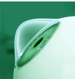 Keratoconus is a non-inflammatory, degenerative disorder of the eye, which causes thinning and bulging of the cornea. This bulging causes distortion of the cornea, and an associated reduction in visual acuity. Corneal scarring can occur in more advanced cases of the disorder leading to further reduction in visual acuity.
Patients with keratoconus report sensitivity to light, streaking and multiple images. The disorder is often detected and diagnosed in adolescent years and achieves its most severe state in the twenties and thirties. If the disorder affects both eyes, the deterioration can greatly impair patients' ability to drive, read and carry out normal everyday tasks. In most cases, corrective lenses can be prescribed to allow patients to function normally. If the disorder progresses to a more advanced state, surgery is an option through either intrastromal corneal ring segments or a corneal transplant amoung other procedures. However, despite the unpredictability of the disease, keratoconus can be successfully treated with little to no infringement on patients' quality of life. State-of-the-art treatments known as cross-linking with riboflavin have shown to restrict the progression of the disease as well.
Keratoconus seems to occur throughout the world affecting 1 in 1000 people and is the most common disorder causing dystrophy of the cornea. The exact cause of keratoconus is unknown but it has been linked with detrimental enzyme activity within the cornea. A genetic link is also apparent, as there is a higher rate of the disorder if family members have been diagnosed with keratoconus and progression of the disease is rapid in Down syndrome patients.
In the beginning, people with keratoconus report merely blurry vision, as the symptoms at the early stages of the disorder are really no different from any other refractive defect of the eye. As the disease advances, vision deteriorates, and in some cases rapidly. Vision becomes impaired at all distances and night vision is very poor making it extremely difficult for patients to drive. In most cases, there is no feeling of pain, however, patients experience sensitivity to bright light, eye strain and itching in the eye. The most common symptom is monocular polyopia, the perception of multiple images or 'ghost' like images. The effect is most clearly noticeable with a high contrast field or light on dark backgrounds. Patients with kerotoconus experience seeing many images of the same point sometimes spread out in disorganized, random patterns. Other symptoms of the disorder are streaking and excessive glowing around light sources.
The first step in diagnosis normally begins with either an ophthalmologist, optometrist or a physician evaluating the patients medical history by assessing previous visual symptoms, ocular diseases and family history. Next, an eye chart is used to determine the visual acuity of the patient and then measurement of the cornea is performed to determine the curvature of the cornea. A measurement of the cornea is normally taken with either a keratometer or retinoscopy depending on the severity and progression of the disease. If keratoconus is suspected further tests may be required, such as a slit lamp examination of the cornea or a keratoscope, to determine if the disease is present by determining the exact topography or curvature of the cornea.
If detected early, keratoconus can be treated with spectacles or soft contact lenses to correct the mild astigmatism which occurs. As the disease advances, spectacles or soft contacts may no longer provide a satisfactory degree of visual acuity for patients, and most optometrists will move to prescribing rigid contact lenses, known as rigid gas-permeables, or RGPs. RGP lenses provide a good level of visual correction, but do not stop the progression of keratoconus. Scleral lenses are sometimes prescribed for cases of advanced or very irregular keratoconus; these lenses cover a greater proportion of the surface of the eye and hence can offer improved stability. However, a soft lens has a tendency to conform to the conical shape of the cornea caused by keratoconus.
The condition will progress in a percentage of patients to a point where vision correction using the above methods will no longer be possible. Surgical options are available. A cornea transplant is one surgical option and keratoconus is the most common ground for this procedure. Penetrating keratoplasty or a cornea transplant, requires the surgeon to graft the donor cornea to the existing eye tissue. The cornea does not have a direct blood supply, so there is no need for a blood type match. The recovery period can take four to six weeks and success rate hovers around 95% making it the most preferred option in stabilizing keratoconus. Other procedures include corneal ring segment inserts, corneal collagen crosslinking with riboflavin, radial keratotomy or LASIK refractive surgery, DALK transplants and Epikeratophakia.
For additional information or to schedule an Eye Examination, please contact us at 1.866.611-7556.
Also Serving:
Sealy, TX - Bellville, TX - Columbus, TX - Katy, TX |